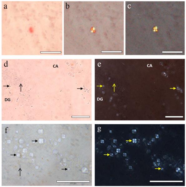
Fig. 2.
Spherulites observed in Alzheimer’s hippocampal tissue stained with Congo red and haematoxylin. a, Congo-red-stained spherulite giving apple-green birefringence under progressively crossed polarisers (a-c), typical of the senile plaques observed in Alzheimer’s disease; d, spherulite structures (solid arrow-heads) in the same tissue section without either the strong affinity for Congo red or e, apple-green birefringence under crossed polarisers. In all sections the unstained spherulites were distributed in the granule cell layer (vertical arrow) of the dentate gyrus (DG) and in a surrounding band in Ammon’s horn (CA) within the pyramidal cell layer; f, hippocampal section with a lighter Congo red stain, haematoxylin-positive cells (vertical arrow) and the spherulites which also notably lack affinity for haematoxylin (solid arrow-heads) shown under partially crossed polarisers; g, same region under fully crossed polarisers. Scale bar in each image is 100 μm.

