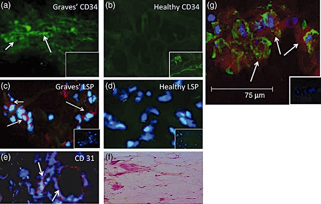
Fig. 1.

CD34+LSP-1+ TSHR+ fibrocytes can be identified in the orbital tissue of patients with TAO but are absent in tissues from healthy donors. (a) CD34 expression (arrows, green FITC) in TAO-derived tissue (inset, negative control staining). (b) Absent CD34 expression in healthy tissue (inset, positive staining control). (c) LSP-1 expression in TAO-derived tissue [red, arrows, nuclei counterstained with DAPI (blue)] (inset negative control). (d) Absence of LSP-1 expression in healthy tissue (inset negative control). (e) CD31 expression in disease-derived tissue is limited to vascular endothelium (red, arrows). (f) H and E stained consecutive thin-sections of the same orbital tissue (40×). (g) Fibrocytes present in orbital tissue from patients with TAO co-express CD34 and TSHR. Nuclei were counterstained with DAPI (blue). Thin sections were then subjected to confocal microscopy. Inset contains a negative staining control. (Reprinted with permission; Douglas, RS et al. Increased generation of fibrocytes in thyroid-associated ophthalmopathy, Copyright 2010, The Endocrine Society.)
