
Fig. 2.
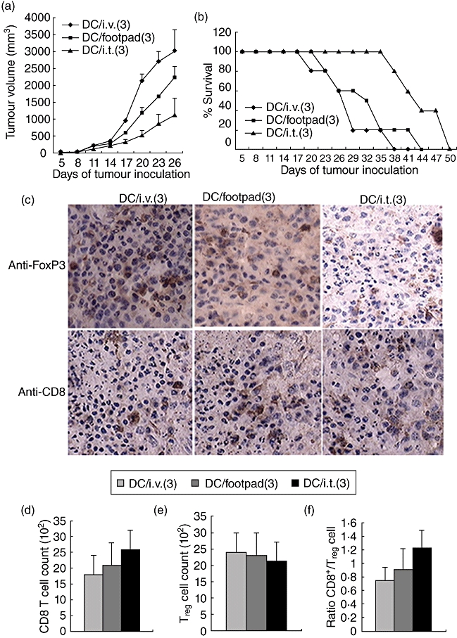
Therapeutic dendritic cells (DCs) fail to reject the established tumours. (a) Tumour growth curve. Mice were inoculated with 0·2 million B16-hepatitis B virus core antigen (HBc) cells subcutaneously. Three days later, mice with tumours at least 3–4 mm in diameter were immunized (intratumorally, intravenously or via footpad) with 1·0 million hepatitis B virus core antigen virus-like particles (HBc-VLP) packaged with cytosine–guanine dinucleotide (CpG) (HBc-VLP/CpG) DCs, and two additional DC vaccinations were administered at 6-day intervals. Lipopolysaccharide (LPS) was injected intraperitoneally directly following each vaccination. DC/ intratumoral (3) means that DCs were injected intratumorally three sequential times. (b) Tumour growth and mouse survival were monitored. (a,b) Representative of three independent experiments (10 mice per group). (c) Tumours were harvested and analysed for the expression of anti-forkhead box P3 (FoxP3) and CD8 by immunohistochemistry. All images were acquired with a 40× objective; 14 days after tumour implantation, tumours were harvested and analysed for the expression of FoxP3 (d) and CD8 (e) by flow cytometry or CD8/FoxP3 (f). (c–f) Cumulative data from three different experiments (three mice per group).