
Fig. 2.
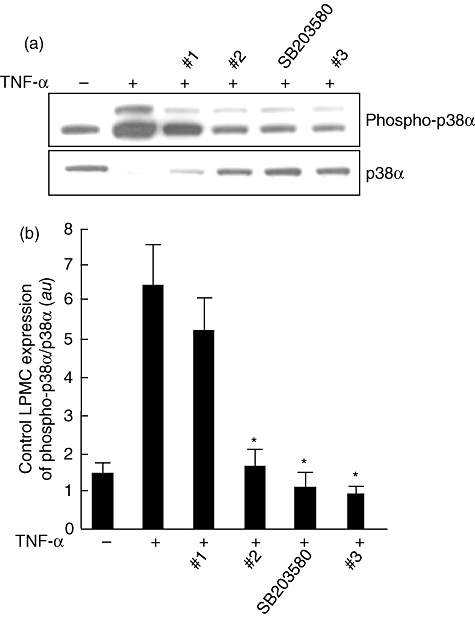
(a) Detection of the phosphorylated form of p38α (phospho-p38α) by immunoblotting in control lamina propria mononuclear cells (LPMCs) cultured with the four p38α inhibitor compounds. LPMCs (1 × 10⁶/ml), isolated from colonic surgical specimens of four control subjects, were incubated for 30 min with or without 10 ng/ml recombinant human tumour necrosis factor (TNF)-α plus p38α inhibitor compounds 1, 2, 3 or SB203580, all at a final concentration of 10 µM. Blots were stripped and analysed for p38α. (b) Densitometry of Western blots. Phospho-p38α expression is normalized for p38α. Results are mean (standard deviation); a.u.: arbitrary units (*P < 0·05 versus cells treated with TNF-α only).