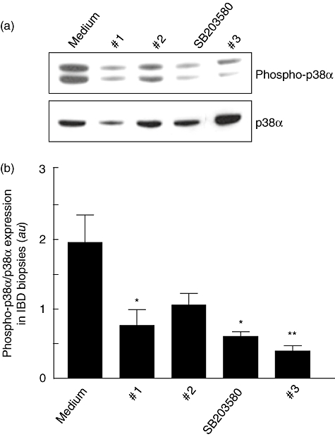
Fig. 4.

(a) Detection of the phosphorylated form of p38α (phospho-p38α) by immunoblotting in biopsy specimens collected from inflamed areas of 14 inflammatory bowel disease (IBD) (seven with Crohn's disease and seven with ulcerative colitis) and cultured for 60 min with p38α inhibitor compounds, namely 1, 2, 3 or SB203580, all at a final concentration of 10 µM. Blots were stripped and analysed for p38α. (b) Densitometry of Western blots. Phospho-p38α expression is normalized for p38α. Results are mean (standard deviation); a.u.: arbitrary units (*P < 0·05 and **P < 0·005 versus cells treated with tumour necrosis factor-α only).
