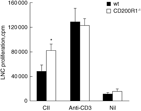
Fig. 3.

Modestly increased T cell proliferation in CD200R1−/− mice. Lymph node single cell suspensions were prepared 14 days after type II collagen immunization and cells were stimulated in the presence of type II collagen or anti-CD3 monoclonal antibody. T cell proliferation was assessed by incorporation of tritiated thymidine. Data are expressed as mean ± standard error of the mean (n = 12) and are representative of two independent experiments. *P < 0·05.
