
Fig. 2.
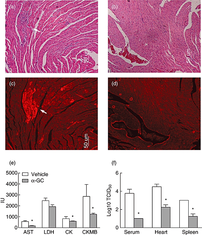
Tissue damage and viral loads of Coxsackievirus B3 (CVB3)-infected mice treated with or without α-galactosylceramide (α-GalCer). BALB/c mice were challenged with CVB3 and treated with α-GalCer (b,d) or vehicle alone (a,c). Samples were taken 7 days after CVB3 challenge. (a,b) Paraffin sections of heart tissue were stained with haematoxylin and eosin (×100). Arrow points to inflammatory cells infiltration. (c,d) Red fluorescence from the same sections and derived Evans blue dye that was taken up by injured cells (arrow) given 4 h before death. (e) Enzyme profiles of CVB3-infected mouse sera. Blood samples were collected at 3 days post-infection. Aspartane aminotransferase (AST), lactate dehydrogenase (LDH), creatine kinase (CK) and creatine kinase muscle band (CKMB) levels were determined as described in Materials and methods. (f) The amount of infectious virus particles were analysed in spleen, heart or serum samples of all animals. Experimental groups consisted of three mice, and experiments were repeated three times. The mean ± standard deviation is shown. Asterisks represent statistical differences between groups. (P < 0·05 as determined by Student's t-test).