
Figure 3. TGF-β and IL-1 stimulation of MMP-9 in CECs is dose-dependent but not isoform dependent.
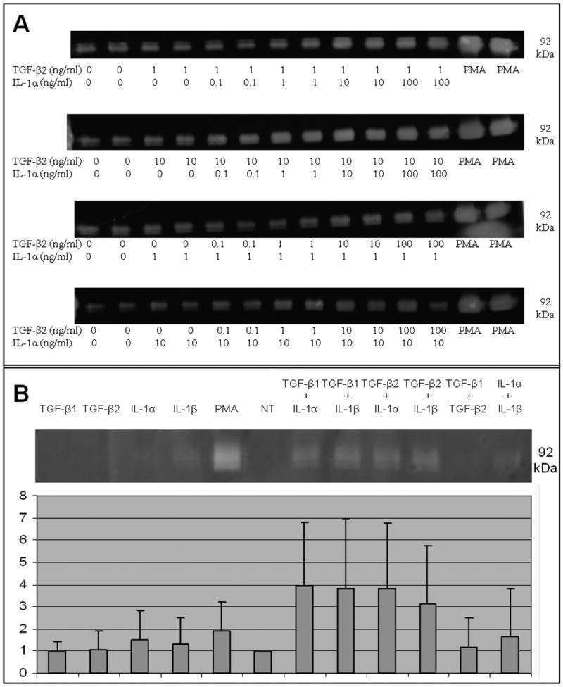
Zymographic analysis of MMP-9 secreted into the media by CECs following cytokine treatment showing (A) TGF-β2 and IL-1β act in a dose-dependent manner to induce MMP9 in CECs. As little as 1 ng of one cytokine combined with 10 ng of the other cytokine is sufficient for stimulation of MMP-9 above basal levels. (B) The stimulation of MMP-9 in CECs by the TGF-β and IL-1 families is not isoform specific; any combination of TGF-β1 or TGF-β2 with either IL-1α or IL-1β is sufficient to up-regulate MMP-9 expression in CECs. Densitometry was performed using the Bio-Rad software “ImageJ”, and MMP-9 stimulation was plotted on a bar graph. Each value is the mean of results obtained from three independent experiments. Error bars indicate standard deviation. ** indicates a p-value of less than 0.005.