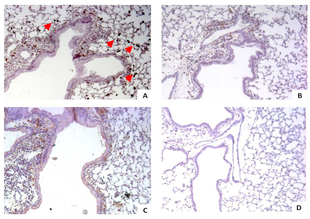
Figure 4. Immunohistochemical staining of Arginase-1 (Arg1).
The four different treatment groups are seen at an original magnification of x100, where Arg1 stains brown. We evaluated the epithelial and subepithelial compartments, of which the latter contains the airway smooth muscle cells, inflammatory cells, and extracellular space. There was no Arg1 staining of airway smooth muscle cells in any of the treatment groups.
A) 6OVA+EtOH. Arg1 stains dark brown and localizes to the subepithelial space and in macrophages (red arrow heads). There is little-to-no Arg1 staining visualized in airway epithelial cells.
B) 6OVA+Sim. Simvastatin treatment (40 mg/kg) of OVA-exposed mice attenuated Arg1 staining in the subepithelial space. No differences were seen compared to 6OVA+EtOH in macrophage Arg1 staining by this qualitative assessment.
C) 6OVA+Sim+MA. Co-treatment with MA (20 mg/kg) partially abrogated the simvastatin effect on Arg1 staining in the subepithelial compartment. No significant differences were seen with respect to macrophage Arg1 staining.
D) FA+PBS. There is no Arg1 staining noted in this air control group.

