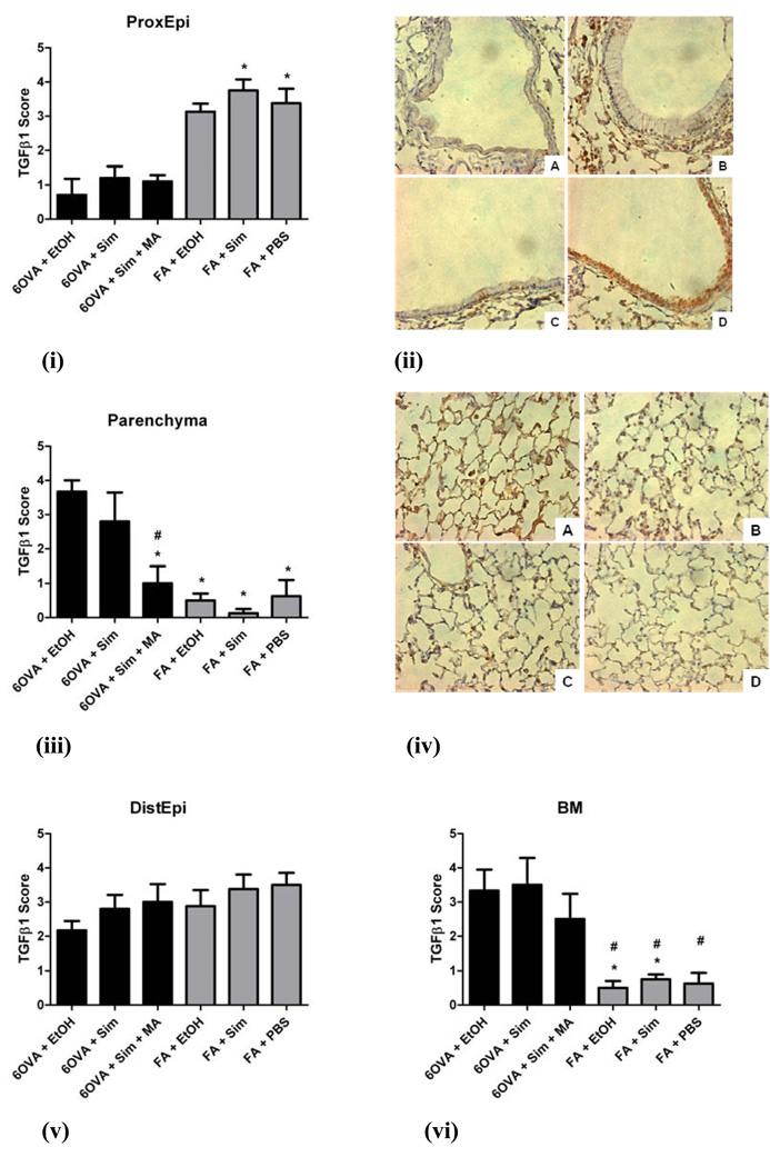
Figure 8. TGFβ1 immunohistochemical staining and scoring in different lung compartments.
The groups illustrated in the histology images are seen at an original magnification of x400, where TGFβ1 stains brown: A) 6OVA+EtOH, B) 6OVA+Sim, C) 6OVA+Sim+MA, and D) FA+PBS. Simvastatin (40 mg/kg) had no statistically significant effect on total TGFβ1 content in the different lung compartments evaluated. Each treatment group had between 4–6 mice. All analyses were done by 1-way ANOVA.
i) In the proximal airway epithelium (ProxEpi), the air controls (FA+Sim, FA+PBS) had greater TGFβ1 content compared to the OVA groups, specifically 6OVA+EtOH (*p<0.05).
ii) Histology corresponding to i) where TGFβ1 stains brown most intensely in the epithelium of the FA+PBS group.
iii) In the lung parenchyma, all three air control groups had much lower TGFβ1 content compared to the 6OVA+EtOH group (*p<0.005). The treatment group 6OVA+Sim+MA showed less TGFβ1 content than the 6OVA+EtOH group (#p<0.005).
iv) Histology corresponding to iii) shows the highest intensity TGFβ1 staining in group A) 6OVA+EtOH, greater than the other OVA groups, and significantly greater than all of the air controls.
v) In the distal airway epithelium (DistEpi), there were no statistically significant differences amongst all six groups (p=NS).
vi) In the basement membrane (BM), the air controls had significantly less TGFβ1 content than the OVA groups, specifically 6OVA+EtOH (*p<0.05) and 6OVA+Sim (#p<0.05).

