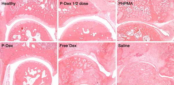
Figure 2.
Light micrographs (magnification, 100x) of ankle joint histology for the six animal groups. Synovial lining and villous hyperplasia, cellular infiltration in periarticular soft tissue, bone and cartilage destruction are clearly evident in the free dexamethasone (Dex), the N-(2-hydroxypropyl)methacrylamide polymer without Dex (PHPMA) and the saline groups. P-Dex, acid-labile N-(2-hydroxypropyl)methacrylamide copolymer-dexamethasone conjugate.

