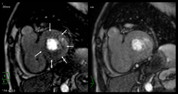
Figure 5.
Early perfusion defect of microvascular impairment without myocardial fibrosis or myocarditis. Perfusion MRI under stress (left figure) showed non-segmental circumferential perfusion defect (arrows). Perfusion MRI at rest (right figure) showed no defects. In this case, delayed enhanced images showed no enhancement (not shown).

