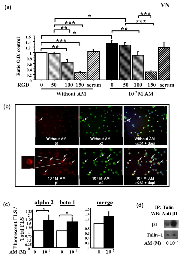
Figure 4.

Adrenomedullin significantly increases cell-surface expression of activated α2 and β1 integrins by rheumatoid fibroblast-like synoviocytes (RA-FLSs). RA-FLSs were incubated with 10-7 M adrenomedullin (AM), or without AM as a control, for 1 hour. Bars indicate mean ± SEM. *P < 0.05; **P < 0.01; ***P < 0.0001. Inhibition adhesion study was done with RGD peptides and scrambled peptide for 30 minutes before AM stimulation. A fluorescence microscope was used for visualization of three sections and image capture. The mean ratio of fluorescent cells from these three sections was calculated as the ratio of the number of fluorescent cells over total cells counted by DAPI staining. Results are given as the ratio of fluorescent cells over control cells without AM and reported as the mean ± SEM of three experiments (three different donors) performed in triplicate. For IP, cell lysates from nonstimulated and AM-stimulated RA-FLSs were immunoprecipitated with anti-talin and then immunoreacted with anti-β1 with Western blot analysis (The figure is representative of two experiments done with two different RA-FLSs). (a) Dose-dependent inhibition of FLSs adhesion by using RGD peptides. Left panel, basal adhesion; right panel, AM stimulation. (b) Observation with fluorescence microscopy of the distribution of activated α2 and β1 integrin expression in RA-FLSs with or without 10-7 M AM. Insert shows rim pattern or cytoplasmic membrane expression of activated integrin. (c) Effect of 10-7 M adrenomedullin on the number of RA-FLSs expressing activated α2 and β1 integrins. (d) IP: Effect of 10-7 M AM on the talin-β1 integrin interaction.
