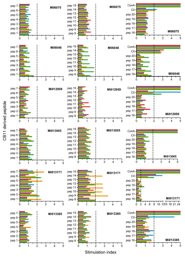
Figure 9.
Fine specificity of proliferating T cells. A detailed analysis of collagen type II (CII)-specific T-cell responses against a cyanogen bromide fragment 11 (CB11)-derived overlapping peptide set (see Table 3) was performed on the day of necropsy with freshly isolated peripheral blood mononuclear cells (PBMC) (green) and cells isolated from the inguinal (red) and axillar (blue) lymph nodes and the spleen (yellow) (animal identification given in the graph). Concanavin (Con) A stimulation was included to establish functional proliferation of the different cell populations and displayed stimulation indices ranging from 25 to 160. In general, CII-specific proliferative responses are better measured in lymph nodes (spleen, axillar and inguinal) than in PBMC. Stimulation index = proliferation of experimental sample (counts per minute)/proliferation of medium control (counts per minute).

