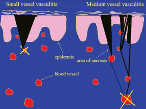
Figure 4. Diagrammatic representation of the difference in small versus medium-sized (deeper) vessel vasculitis and the impact on skin findings.
A superficial vasculitis (left side of diagram) leads to a wedge-shaped area of necrosis and, thus, a well defined and regular skin purpura or necrosis. Conversely, occlusion of a deep vessel (right side of diagram) leaves open the chance for anastomosing vessels to alter the effect at the skin.

