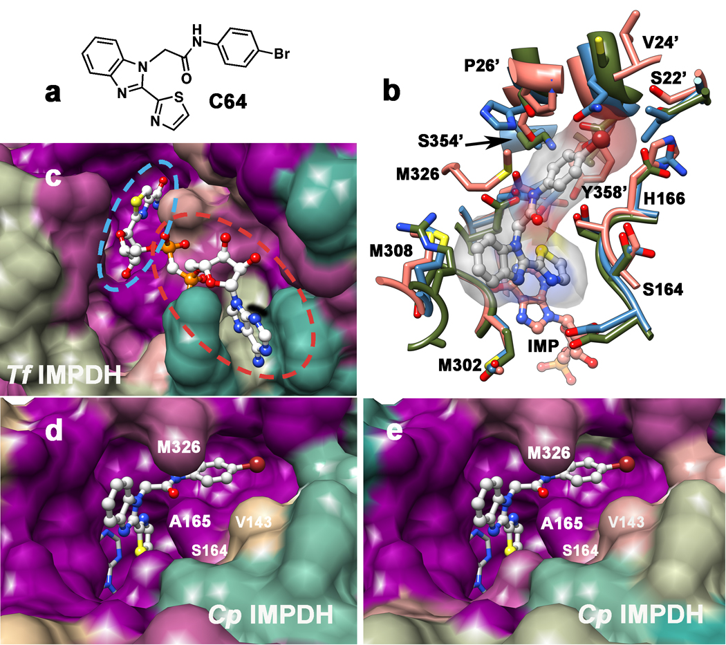
Figure 2. Structures of IMPDH inhibitor binding sites.
a. Structure of C64. b. Structure of the CpIMPDH•IMP•C64 complex (salmon; PDB accession 3KHJ (MacPherson, et al.)) and resistant IMPDHs from T. foetus (green; 1LRT (Gan, et al., 2002)) and Chinese hamster (blue; 1JR1, nearly identical to human IMPDH2 (Sintchak, et al., 1996)). Residues within 5 Å of C64 are displayed. C64 is shown in gray with a transparent surface; CpIMPDH residues are labeled; residue from the adjacent monomer are denoted with a '. Residues Thr221 and Glu329 are hidden under C64. c. The surface of the NAD binding site rendered by conservation of residues in prokaryotic IMPDHs from T. foetus, C. parvum, H. pylori, B. burgodorferi, S. pyogenes and EcIMPDH. Dark magenta, 100% conserved; tan, 63%; dark cyan, 25%. The NAD analog tiazofurin adenine dinucleotide (TAD) is light gray. The tiazofurin binding site is circled in blue and the ADP binding site is circled in red. d. The surface of the C64 binding site rendered by conservation of residues in sensitive IMPDHs from C. parvum, H. pylori, and B. burgdorferi, as well as the S250A/L444Y variant of EcIMPDH. Ser164, Met326 and Ser354 are within 5 Å of C64; Val143 is 8 Å away. Dark magenta, 100% conserved; tan, 60%; dark cyan, 20%. C64 depicted in ball-and-stick in light gray and IMP is shown in stick in dark gray. e. The surface of the C64 binding site rendered by conservation of residues in CpIMPDH and IMPDHs from 19 pathogenic bacteria: Acinetobacter baumannii (wound infection), Bacillus anthracis (anthrax), Bacteroides fragilis (peritoneal infections), Brucella abortus/melitensis/suis (brucellosis), B. burgdorferi (Lyme disease), Burkholderia cenocepacia (infection in cystic fibrosis), Bu. mallei (glanders), Bu. pseudomallei (melioidosis), Campylobacter jejuni (food poisoning), C. lari (food poisoning), Coxiella burnetii (Q fever), Francisella tularensis (tularemia), H. pylori (gastric ulcer/stomach cancer), Listeria monocytogenes (listeriosis), Staphylococcus aureus (major cause of nosocomial infection), S. pyogenes (major cause of nosocomial infections) and S. pneumoniae (pneumonia). Dark magenta, 100% conserved; tan, 63%; dark cyan, 25%. Alignments were constructed with CLUSTALW2 and molecular graphics images were produced using the UCSF Chimera package from the Resource for Biocomputing, Visualization, and Informatics at the University of California, San Francisco (supported by NIH P41 RR-01081) (Pettersen, et al., 2004). Supported by Figure S3.

