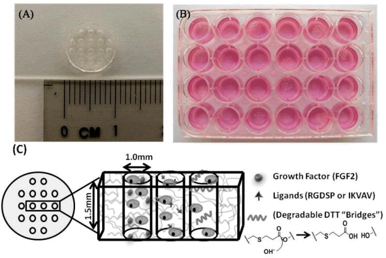
Figure 1.

A) 8kDa PEG hydrogel array “background” showing an array of 16 cylindrical spots. B) Image demonstrating 24 hydrogel arrays within 24-well tissue culture plate. C) Schematic demonstrating incorporation of stem cells, growth factors, peptide ligands, and hydrolytically degradable DTT “bridges” into hydrogel array spots.
