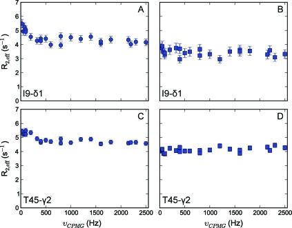
Figure 4.
Representative methyl transverse 1H relaxation dispersion profiles for Ile-δ1 and Thr methyl groups for calbindin D9k recorded at 600 MHz. Panels A and C show spurious dispersion profiles originating from homonuclear scalar coupling; in panels B and D, the source of this artifact has been removed in the NMR experiment.

