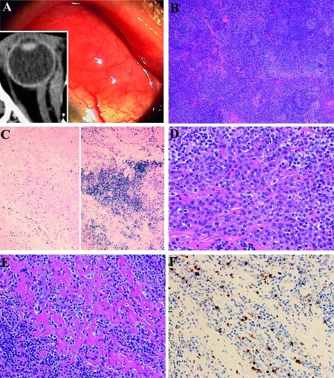
Figure 2.
Histological and immunohistochemical findings in conjunctival marginal zone B cell lymphoma with IgG4-positive plasma cells from patient 6. (A) Salmon-pink mass lesions on the left bulbar conjunctiva. CT image shows medial rectus muscle enlargement (insert). (B) Histological study shows dense lymphocytic proliferation with sclerosis and follicles. H&E staining, original magnification ×40. (C) Immunostaining for λ (left) and κ (right) shows light-chain restriction. Southern blot analysis shows immunoglobulin heavy-chain gene rearrangement. In situ hybridisation, original magnification ×40. (D) Higher magnification shows small and medium-sized lymphocytes with plasma cells and sclerosis. H&E staining, original magnification ×400. (E) Another higher magnification shows lymphoplasmacytic infiltrations with abundant sclerosing lesions. H&E staining, original magnification ×400. (F) Immunostaining for IgG4 reveals IgG4-positive plasma cells in the lymphoplasmacytic lesions. The IgG4:IgG ratio was 0.53. Immunoperoxidase staining, original magnification ×400.

