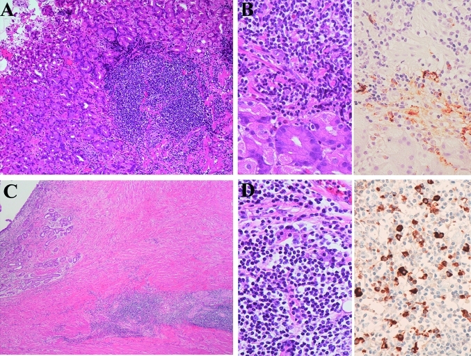
Figure 4.
Gastric biopsy samples of patients 2 and 3. (A) Histology of patient 2 shows lymphoplasmacytic proliferations in the stomach. H&E staining, original magnification ×100. Immunostaining did not show light-chain restriction; however, the analysis of the specimen on PCR showed immunoglobulin heavy-chain gene rearrangement. (B) Higher magnification (left) shows lymphoplasmacytic infiltrations. H&E staining, original magnification ×400. Immunostaining for IgG4 (right) shows IgG4-positive plasma cells. The IgG4:IgG ratio was 0.67. Immunoperoxidase staining, original magnification ×400. (C) Histology of patient 3 shows lymphoplasmacytic proliferations. H&E staining, original magnification ×100. Immunostaining did not show light-chain restriction; however, the analysis of the specimen on PCR showed immunoglobulin heavy-chain gene rearrangement. (D) Higher magnification (left) shows lymphoplasmacytic infiltrations. H&E staining, original magnification ×400. Immunostaining for IgG4 (right) showed IgG4-positive plasma cells. The IgG4:IgG ratio was 0.70. Immunoperoxidase staining, original magnification ×400.

