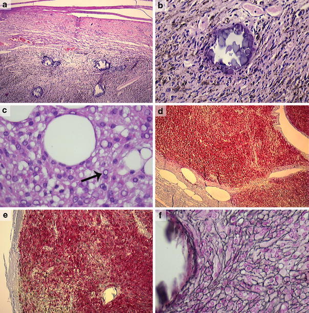
Fig. 1.

Melanotic schwannoma (case 4) showing a circumscribed tumor, covered by a fibrous capsule and containing several psammoma bodies (a) (×50). Detail of the same lesion showing spindle cell morphology, an ample amount of melanin pigment and a psammoma body (case 4) (b) (×200). Melanotic schwannoma (case 8) with more epithelioid cell morphology, nuclear pseudoinclusions (arrow) and large vacuoles resembling fat (c) (×400). Strong and diffuse positivity for, respectively, S-100 and HMB-45 in a melanotic schwannoma (case 4) (d, e) (×25, ×100). Same lesion with pericellular deposition of basement membrane material (case 4) (f) (Laguesse stain, ×400)
