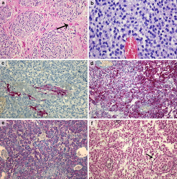
Fig. 2.

Intermediate-grade melanocytoma (case 20) with invasion of neuroglial tissue; note the Rosenthal fibers (arrow) in the surrounding neuropil (a) (×200). Epithelioid cell morphology with bland nuclei and small nucleoli in a melanocytoma (case 22) (b) (×400). Focal positivity for S-100 in the same lesion (case 22) (c) (×200). Diffuse positivity for HMB-45 and Melan-A, respectively, in the same lesion (d, e) and a nested staining pattern in the reticulin stain (f) (case 22) (Laguesse stain, ×200)
