Fig. 3.

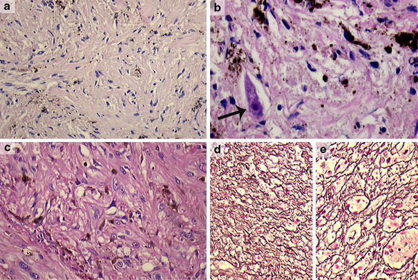
Case 24 was a variably, partly heavily pigmented lesion consisting of spindle to pleomorphic cells (a, c) (×200), showing infiltrative growth in a ganglion at the lumbar spinal region with dispersed incorporated ganglion cells (arrow) (b) (×400). The nuclei showed marked atypia with conspicuous nucleoli (c) (×200). In the reticulin stain, both a pericellular (d) and nested staining pattern were seen (e) (Laguesse, ×100). We, therefore, favored the diagnosis of a melanotic schwannoma with atypical features suggestive of aggressive behavior. Molecular analysis of this case did not reveal mutations in the GNAQ gene
