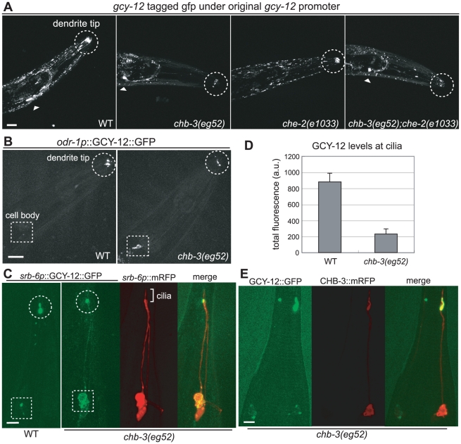
Figure 4. chb-3 is required for proper localization of the GCY-12 guanylyl cyclase to cilia.
(A) Expression of GFP-tagged, full-length GCY-12 under the control of the native promoter (GCY-12::GFP). Expression of the ttx-3p::mRFP injection marker in the AIY neurons is also shown (arrowheads). (B) Expression of GFP-tagged, full-length GCY-12 under the control of the odr-1 promoter (odr-1p::GCY-12::GFP). (C) Expression of GFP-tagged, full-length GCY-12 under the control of the srb-6 promoter (srb-6p::GCY-12::GFP). srb-6p::mRFP was also expressed to visualize the morphology of the cilia in chb-3(eg52). (D) Ciliary GFP levels in animals expressing odr-1p::GCY-12::GFP were quantified using the confocal 3D analysis. Numbers of examined animals; wild-type (16), chb-3 (18). Error bars indicate s.e.m. (E) Decreased GCY-12 levels in the cilia of chb-3(eg52) were rescued cell autonomously. The tail region of a chb-3(eg52) animal carrying two extrachromosomal transgenes, Ex[srb-6p::GCY-12::GFP] and Ex[srb-6p::CHB-3::mRFP], was shown. In this animal, Ex[srb-6p::CHB-3::mRFP] showed mosaic expression only in the right phasmid neuron, and only the right phasmid ciliary level of GCY-12::GFP was increased. (A, B, C, E) Projection of confocal microscopic sectioning images. Dendrite tips and cell bodies are marked with circles and squares, respectively. Head (A, B) and tail (C, E) regions of young adults are shown. Scale bars represent 20 µm (A, B) and 5 µm (C, E).

