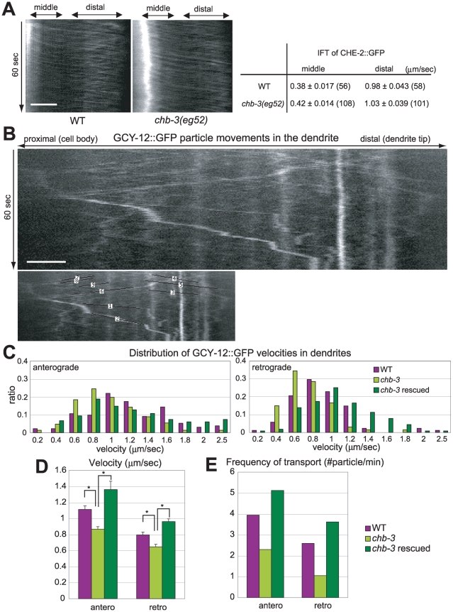
Figure 6. chb-3 is required for proper GCY-12::GFP transport along the dendrites to cilia.
(A) left; Kymographs depicting CHE-2::GFP particle motility within the cilia. right; The velocities of CHE-2::GFP particles along the middle segments and the distal segments of cilia are shown as the average ± s.e.m. (number of particles). (B) A kymograph depicting GCY-12::GFP particle motility along the dendrite of a phasmid sensory neuron in a wild-type animal. Corresponding lines, with which the velocity and the frequency were analyzed, are also shown. The movie is available upon requests. (A, B) The horizontal and vertical axes represent distance and time, respectively. Horizontal scale bar; 2 µm. (C) Distribution of GCY-12::GFP particle velocities in dendrites. (D) Average velocities of GCY-12::GFP particles in anterograde and retrograde directions in the dendrites with s.e.m. Asterisk indicates the significant difference at p<0.001 (t test). (E) Frequencies of GCY-12::GFP particles in anterograde and retrograde directions in the dendrites. (C, D, E) Wild-type, chb-3(eg52) and chb-3(eg52);Ex[srb-6p::CHB-3::mRFP](chb-3 rescued) animals were carrying Ex[srb-6p::GCY-12::GFP] transgene, and their dendrites of the phasmid neurons were observed. Total time of observation, total numbers of particles in anterograde direction and in retrograde direction are follows; WT (34 min, 134, 88), chb-3 (64 min, 147, 67), chb-3 rescued (32 min, 164, 116).

