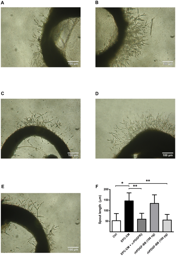
Figure 3. Angiogenic potential of EPC-CM on ex vivo aortic ring assays.
Incubation with EPC-CM (B) enhanced the formation of vascular outgrowth from 1 mm rat aortic ring embedded in growth factor reduced-Matrigel™ compared to control medium incubation (A). This enhanced EC cord structure outgrowth could be blocked by the addition of 1 µg/ml PDGFRβ antibody into EPC-CM (C). A similar vascular sprouting extent could only be observed by stimulation aortic ring with 100 ng/ml rhPDGF-BB (1000-times concentrated than the content in EPC-CM) (D), but not with the concentration at 100 pg/ml (E). The extents of vascular outgrowth were quantitatively analyzed and presented by the length of the sprouts (D). *, #, P<0.001.

