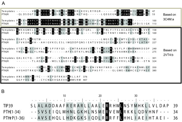
Figure 1.
Alignments. Black shaded residues are identical and grey shaded residues are similar according to PAM250 similarity matrix. A: Alignment between PTH2R and the templates used for homology modelling; thick border box - extracellular domain; thin border box - transmembrane region; no box - intracellular domain. B: Alignment of tuberoinfundibular peptide of 39 residues (TIP39), parathyroid hormone (PTH) and parathyroid hormone related protein (PTHrP). The amphipatic nature of the peptides is visible through the pattern of conserved hydrophobic residues in the C-terminal portion of the alignment.

