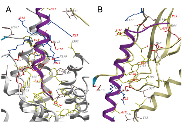
Figure 3.
Close-up of the binding region between PTH2R and the tuberoinfundibular peptide of 39 residues. The extracellular domain (ECD) in khaki, regions without template in blue, the TM region in white and the tuberoinfundibular peptide of 39 residues (TIP39) in magenta. Residues in the binding interface of TIP39 and the receptor (within 3Å distance of each other) are shown in sticks. Acidic residues are coloured red, polar residues are coloured pink, basic residues are coloured blue and hydrophobic residues are coloured yellow. Residues in TIP39 are labelled in red and residues in the receptor are labelled in gray. A. Interactions of the N-terminus of TIP39 with the TM region. B. Interactions of the C-terminus of TIP39 with the ECD.

