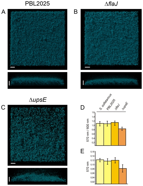
Figure 7. Three day matured static biofilms of S. solfataricus PBL2025, ΔflaJ and ΔupsE.
Biofilms of PBL2025, ΔflaJ and ΔupsE were stained with DAPI and analyzed by CLSM (A–C, respectively). Complementary, a microtitre plate assay was performed for 72 hrs with all three strains and biofilm formation is presented in D as the correlation of the crystal violet absorbance (OD570 nm) divided by the optical density of the planktonic cells (OD600 nm) and in E the crystal violet absorbance (OD570 nm) is indicated. Bars are 20 µm in lengt.

