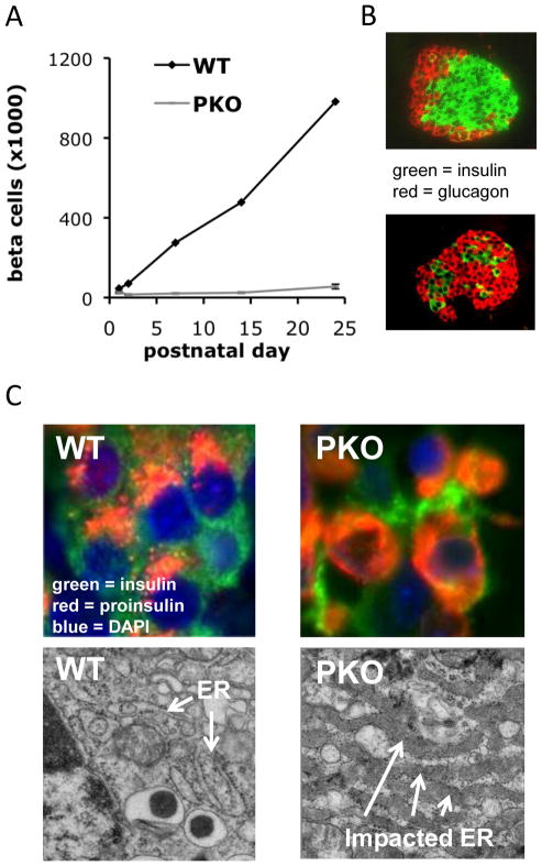
Figure 2.
PERK deficiency results in diminished proliferation of beta cells and a severe defect in proinsulin trafficking. (a) Beta cell mass normally increases 20–40 fold during the first four postnatal weeks in mice (24). In contrast, beta cell mass increases only about 2-fold in Perk KO mice. (b) Images of wildtype and Perk KO islets at the 3rd postnatal week are shown. (c) Proinsulin (red) normally accumulates in the ER-Golgi Intermediate Compartment and Golgi adjacent to the nucleus (blue) whereas insulin (green) is dispersed through the cytoplasm. In a large fraction of beta cells in Perk KO (PKO) mice, proinsulin accumulates in the ER and is not trafficked to the Golgi (24). (c) The ER in wildtype beta cells exhibits an elongated, tubular structure dotted with ribosomes, whereas the ER in Perk KO mice is highly distended with high electron density due to the accumulation of a large amount of proinsulin and other client proteins.

