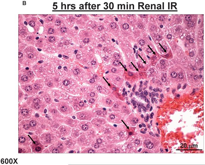
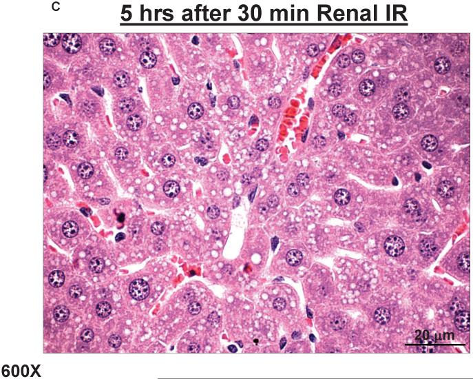
Figure 2. Acute hepatic injury with increased hepatic necrosis and vacuolization after ischemic AKI.
Representative photomicrographs of liver from 6 experiments (hematoxylin and eosin staining, magnification 600X) of mice subjected to sham-operation (Sham) or to 30 min. renal ischemia and 5 hrs of reperfusion (30 min. renal IR). Sham operated animals show normal-appearing hepatocyte parenchyma (A). Five hours after 30 min. renal IR (B, C), nuclear and cytoplasmic degenerative changes, centrilobular necrosis (B, arrows), marked hepatocyte vacuolization (C) and congestion were observed.


