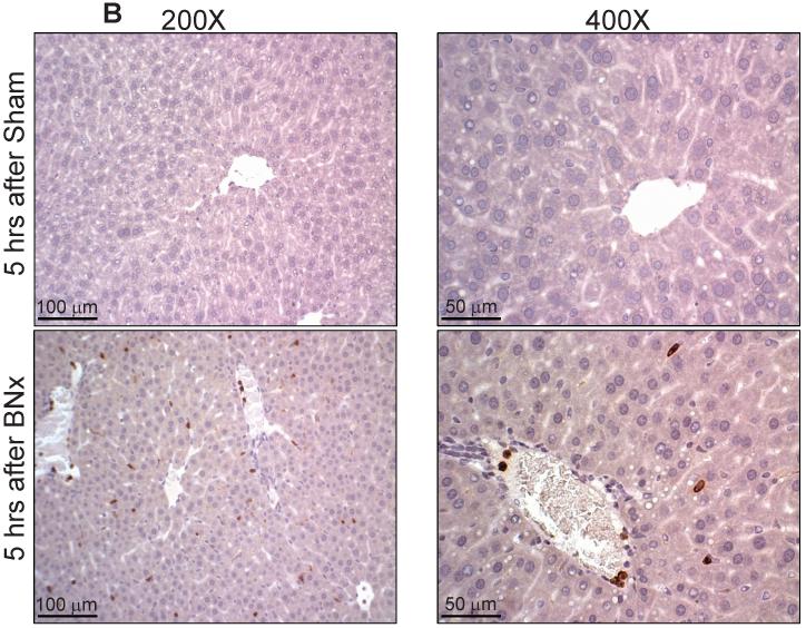
Figure 3. Increased hepatic inflammation after ischemic or non-ischemic AKI.
A. Representative gel images and band intensity quantifications of semi-quantitative RT-PCR of the pro-inflammatory markers ICAM-1, TNF-α, IL-6, IL-17A, KC, MCP-1 and MIP-2 from liver tissues of mice subjected to sham-operation (Sham), bilateral nephrectomy (BNx) or 30 min. renal IR (RIR). Liver tissues were harvested 5 hrs after sham-operation or AKI induction. *P<0.05 vs. sham-operated mice. Error bars represent 1 SEM. B. Representative photomicrographs (200X and 400X) of 4 experiments of immunohistochemistry for neutrophil infiltration (dark brown stain) in the liver tissues harvested from mice subjected to sham-operation (Sham) or to bilateral nephrectomy (BNx) 5 hrs prior.


