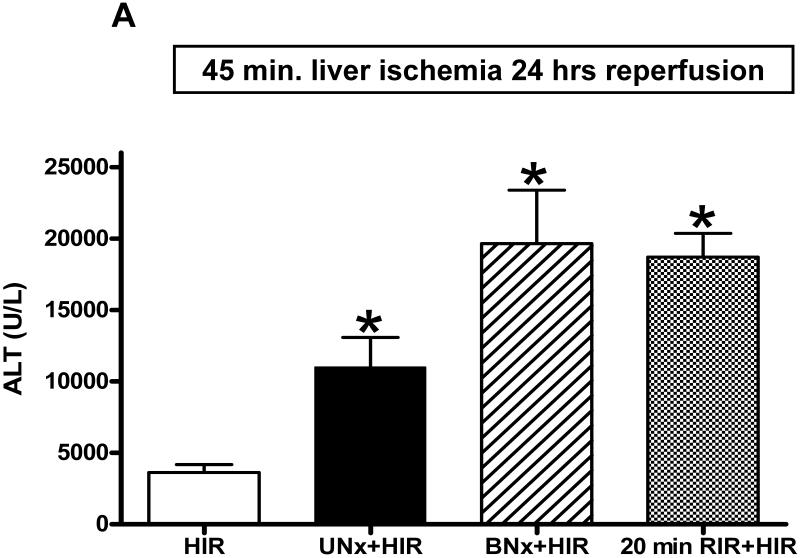
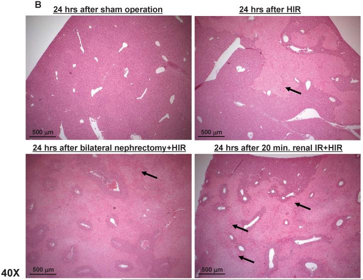
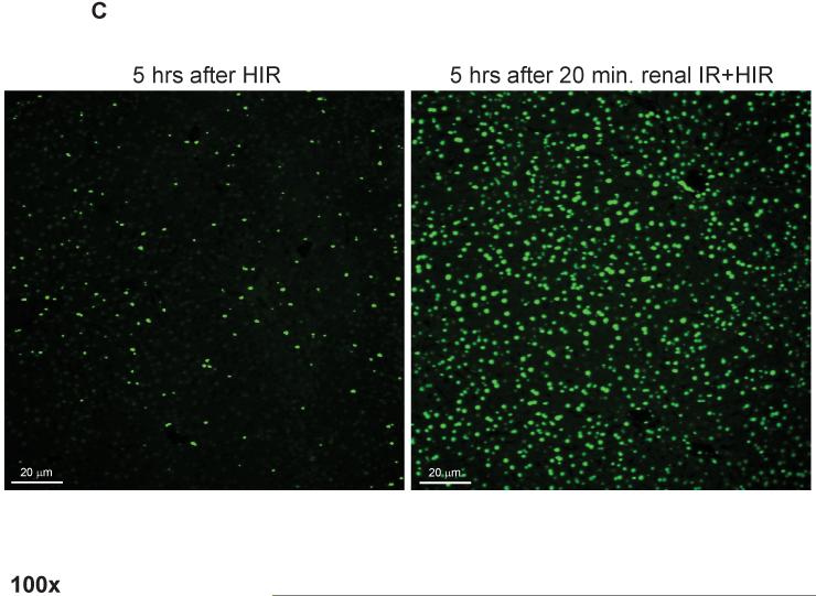
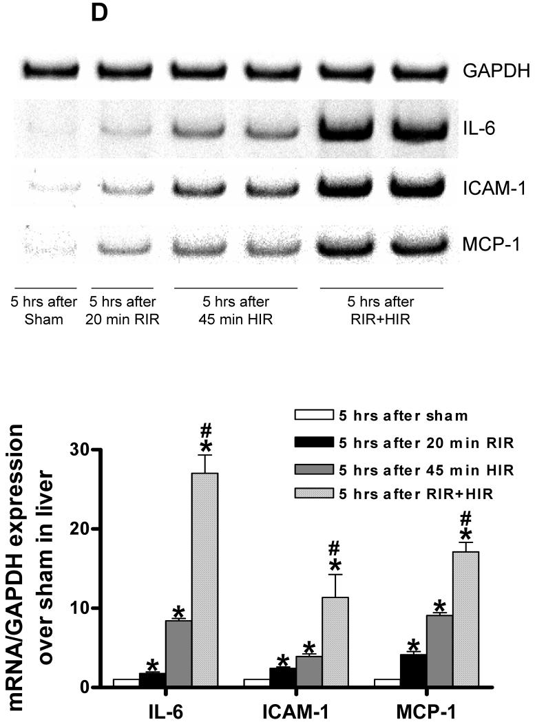
Figure 5. Exacerbated hepatic IR injury (necrosis, apoptosis and inflammation) after ischemic or non-ischemic AKI.
A. Plasma alanine aminotransferase (ALT) levels in mice subjected to sham-operation (Sham, N=4), 45 min. hepatic ischemia and reperfusion (HIR, N=6), HIR coupled with unilateral nephrectomy (UNx+HIR, N=6), HIR coupled with bilateral nephrectomy (BNx+HIR, N=6) or HIR coupled with 20 min. renal ischemia and reperfusion (RIR+HIR, N=8) 24 hrs prior. *P<0.05 vs. HIR mice. Error bars represent 1 SEM. B. Representative hematoxylin and eosin staining photomicrographs in liver sections (magnification X40) in mice subjected to sham-operation, 45 min. hepatic ischemia and reperfusion (HIR), HIR coupled with bilateral nephrectomy or HIR coupled with 20 min. renal ischemia and reperfusion 24 hrs prior. Necrotic hepatic tissue appears as light pink. Arrows indicate vascular congestion and inflammation. C. Representative fluorescence photomicrographs (of 4 experiments) of liver sections illustrating apoptotic nuclei [terminal deoxynucleotidyl transferase biotin-dUTP nick end-labeling (TUNEL) fluorescence staining, 100X] from mice subjected to 45 min. hepatic ischemia and reperfusion (HIR) or HIR coupled with 20 min. renal ischemia and reperfusion 24 hrs prior. D. Representative gel images (top) and band intensity quantifications (bottom) of semi-quantitative RT-PCR of the pro-inflammatory markers ICAM-1, IL-6 and MCP-1 from liver tissues of mice subjected to sham-operation (Sham), 20 min. renal ischemia and reperfusion (RIR), 45 min hepatic ischemia and reperfusion (HIR) or 20 min RIR plus HIR. Liver tissues were harvested 5 hrs after sham-operation or AKI induction. *P<0.05 vs. sham-operated mice. Error bars represent 1 SEM. #P<0.01 vs. HIR mice.


