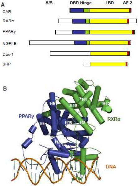
Fig. 1. Domain organization of nuclear receptors.
A, A schematic representation showing functional domains of different nuclear receptors. The DBD is labeled in blue, the hinge is in green and the LBD is in yellow. The presence of AF-2 is indicated in red. B, Multi-domain structure of the PPARγ/RXR/DNA complex in ribbon representation. The crystal structures of PPARγ(blue) and RXRα(green) heterodimer (top) on PPAR response DNA sequence (Bottom).

