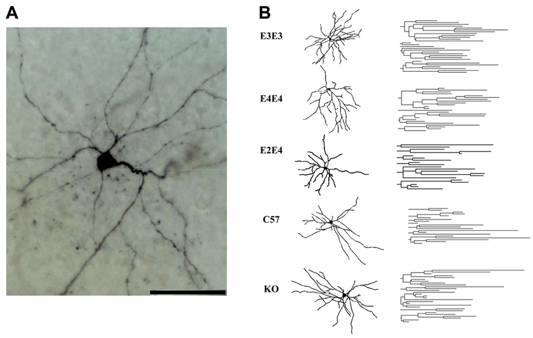
Figure 2.
A. High power image of a representative pyramidal-like lateral amygdala neuron from an apoE3 TR mouse at 7 months of age (scale bar, 50 µm). B. Camera lucida reconstructions (left) and dendrograms (right) corresponding to representative neurons from the indicated cohorts. Quantitative morphological analysis is presented in Table 2.

