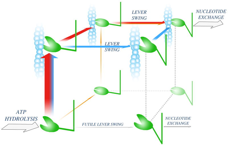
Figure 2. Powerstroke pathways.
Following ATP hydrolysis, the myosin head adopts a weak actin-binding (open-cleft), up-lever state (lower left corner). Three possible pathways leading to an effective powerstroke are shown as orange, red and blue arrows. On the orange pathway, cleft closure is followed by association to actin and subsequent lever swing. The other two pathways (red and blue) start with actin attachment. On the red pathway, cleft closure precedes lever swing. On the blue pathway, lever swing occurs while the cleft is still open, and the pathway is completed with cleft closure. Experimental data indicate that the flux through the orange pathway is limited, whereas the red and blue pathways might both convey significant fluxes. A futile lever swing, which would lead to an ATP-wasting cycle (grey arrow), is kinetically blocked (Box 3).

