Abstract
Animal models are widely used to study cartilage degeneration. Experimental interventions to alter contact mechanics in articular joints may also affect the loads borne by the leg during gait and consequently affect the overall loading experienced in the joint. In this study, force plate analyses were utilized to measure parameters of gait in the rear legs of adult rats following application of a varus loading device that altered loading in the knee. Adult rats were assigned to Control, Sham, or Loaded groups (n≥4/each). Varus loading devices were surgically attached to rats in the Sham and Loaded groups. In the Loaded group, this device applied a controlled compressive overload to the medial compartment of the knee during periods of engagement. Peak ground reaction forces during walking were recorded for each rear leg of each group. Analyses of variance were used to compare outcomes across groups (Control, Sham, and Loaded), leg (Contralateral, Experimental) and device status (Disengaged, Engaged) to determine the effects of surgically attaching the device and applying a compressive overload to the joint with the device. The mean peak vertical force in the experimental leg was reduced 30% in the Sham group in comparison to the contralateral leg and the Control group indicating an effect of attaching the device to the leg (p<0.01). No differences were found in ground reaction forces between the Sham and Loaded groups with application of compressive overloads with the device. The significant reduction in vertical force due to the surgical attachment of the varus loading device must be considered and accounted for in future studies.
Keywords: Gait, animal model
1. INTRODUCTION
Animal models of cartilage degeneration associated with osteoarthritis employ experimental interventions to alter contact mechanics in articular joints. These interventions may also affect the load borne by the experimental leg during gait and subsequently affect the overall load experienced in the joint (O'Connor et al., 1989; Vilensky et al., 1994). Small animal models involving ligament transection or menisectomy are used widely to study cartilage degeneration. However, little is known regarding the effects of these commonly used procedures on the resulting joint contact stresses. In the development of new models to study the contribution of load alteration in the development of cartilage degeneration, it is important to characterize any effects that the interventions have on gait as changes in ground reaction forces will result in changes in joint forces. This is important for establishing how the experimental intervention replicates normal and diseased human conditions.
The present study examines gait alterations in the rat following surgical attachment of a varus loading device (VLD) (Figure 1) and subsequent application of a compressive overload to the medial compartment of the knee utilizing the device. The VLD was previously developed and applied to small animals to study the effects of chronic altered loading on articular cartilage of the knee (Roemhildt et al., 2009; Roemhildt et al., 2010(Roemhildt et al., 2009). Engaging the VLD applies a static varus moment about the knee resulting in a controlled compressive overload of the medial compartment and an equivalent decrease in load to the lateral compartment without constraining the joint or inhibiting normal range of motion. This static overload is in addition to the normal static and dynamic loads in the joint. The magnitude of the overload can be controlled by setting the torque of the torsion spring. When the device is disengaged, no load alteration is applied to the knee.
Figure 1.
(A) Oblique view of varus loading device (VLD) applied to rat femur and tibia via transcutaneous bone plates secured with bone screws. The axis of the bearing is aligned with the epicondylar axis of the femur utilizing fluoroscopy. Setting the torque of the torsion spring applies a varus moment to the distal tibia which results in an increased compressive load in the medial compartment of the tibiofemoral joint. (B) VLD disengaged. To disengage the VLD the offset link and a portion of the load link are removed and the remaining load link is rotated into alignment with the femur tube and secured, thus removing the varus moment. (C) Anterior-posterior view of rat femur and tibia with a VLD attached and engaged illustrating the delivery of a compressive overload to the medial (M) compartment and a decrease in loading in the lateral (L) compartment. (D) Rat with VLD attached and engaged.
The objectives of the current study were 1) to evaluate the effect of surgically attaching the VLD to the rat hind limb on contact time and ground reaction forces during walking, and 2) to evaluate the effect of applying a compressive overload to the medial compartment of the knee via the VLD on these gait parameters.
2. MATERIALS AND METHODS
Following approval by the Institutional Animal Care and Use Committee, 13 Sprague-Dawley rats greater than 9 months of age (mean weight ± standard deviation: 6.47±0.26 N) were randomly assigned to Control (n=5), Sham (n=4), or Loaded groups (n=4). Rats in the Sham and Loaded groups underwent surgery to attach transcutaneous bone plates to the lateral aspect of the left tibia and femur. The fascia between the biceps femoris, vastus lateralis, and superficial gluteal muscle was bluntly dissected to expose the lateral aspect of the femur. The fascia between the medial tibialis cranialis and the medial peroneus longus muscles was bluntly dissected to expose the tibia. Bone plates were attached with 1-mm diameter transcortical bone screws. Following a 2-week recovery, these rats were fit with a VLD which was engaged to apply a compressive overload to the medial compartment 12 hours per day for 12 weeks (Figure 1). By close observation, all rats ambulated normally throughout the study. The magnitudes of compressive overload applied to the medial compartment of the knee were 0% body weight (BW) for the Sham group or either 50% (n=1) or 80% BW (n=3) for the Loaded group.
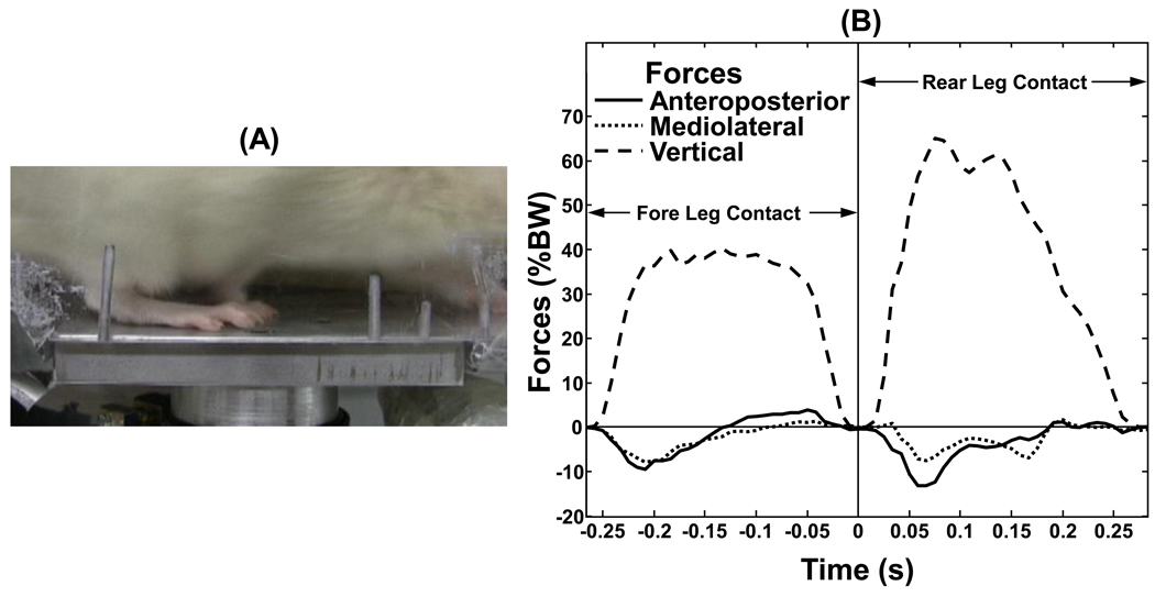
Methods of Zumwalt et al. were adapted to measure ground reaction forces in the rear legs of rats during walking (Zumwalt et al., 2006). The testing apparatus consisted of an acrylic chute approximately 114 (length) × 23 (width) × 40 (height) cm, a 6 DOF load cell (20E12A-I25, JR3 Inc., Woodland, CA, USA) placed at the center of the length and outer edge of the chute (force plate 6.35 (width) × 10 (length) cm), and video cameras positioned laterally and longitudinally. Video recordings acquired at 60 frames per second were used to confirm gait, rear foot contact with the force plate, and measure the speed at which the rat crossed the force plate. Rats were acclimated to the testing chute and protocol through multiple weekly training sessions for 2–3 weeks prior to testing. Data were collected from 10 passes across the force plate for each rear leg (contralateral and experimental) of each rat during spontaneous walking during weeks 10–12 of loading. Vertical, anteroposterior (braking and propulsive) and mediolateral forces were sampled at 120 Hz and normalized by BW (Figure 2). Contact time was determined from the start to end of the rear foot ground contact as determined from the vertical forces. Prior to statistical analysis, data were screened to ensure a consistent walking gait, speed > 100 mm/s and that only the rear foot landed solely on the force plate (Table 1).
Figure 2.
(A) Image of rat crossing the force plate illustrating isolated rear foot contact. (B) Anteroposterior (braking: −, propulsive: +), mediolateral (lateral: +, medial: −) and vertical gait force time histories as a percent of body weight (% BW) of a single trial. Contact time is the time the rear leg is in contact with the force plate as measured by the vertical force.
Table 1.
Distribution of rats and collected data for the experimental and contralateral limbs of animals in the Control, Sham and Loaded groups. For Engaged trials, the attached varus loading device (VLD) was engaged which applied a compressive overload to the medial compartment of the knee. For Disengaged trials, the attached VLD was disengaged so that no overload was applied. Note that if there is no binding in the device, there should be no differences between having the VLD engaged or disengaged for the Sham group since there is no additional loading applied.
| Group | # of Rats | Total # of Trials |
Total Contralateral |
Total Experimental |
Contralateral Disengaged |
Contralateral Engaged |
Experimental Disengaged |
Experimental Engaged |
|---|---|---|---|---|---|---|---|---|
| Control | 5 | 54 | 23 | 31 | NA | NA | NA | NA |
| Sham | 4 | 87 | 41 | 46 | 23 | 18 | 23 | 23 |
| Loaded | 4 | 58 | 25 | 33 | 14 | 11 | 20 | 13 |
Two sets of statistical analyses were performed to address study objectives. First, to examine the effect of surgically attaching the VLD to the limb, comparisons were made between Control and Sham groups. For the Sham group, only trials with the VLD disengaged were used in order to evaluate differences across groups independent of any additional confound. These analyses were based on two-way repeated measures analyses of variance with group (Control, Sham) as an across-animal factor and leg (Contralateral, Experimental) as a within-animal factor. Secondly, to examine the effects of application of the compressive overload by the VLD, three-way repeated measures analyses of variance were performed. The model included the across-animal factor, group (Sham, Loaded) and within-animal factors, VLD status (Disengaged, Engaged) and leg (Contralateral, Experimental). Outcome measures tested were contact time and vertical, braking, propulsive, medial and lateral forces. Replicate trials were averaged for each animal within each experimental condition prior to analysis. All analyses were performed using SAS statistical software (SAS Institute, Cary, NC) with statistical significance determined based on α = 0.05.
3. RESULTS
3.1 Attachment of the VLD
In analyses across Control and Sham groups to examine the effect of surgical attachment of the VLD, the mean peak vertical force was reduced 30% in the experimental limb of Sham animals (p<0.01) relative to the same limb of the Control group and relative to the Contralateral limb within the Sham group (Figure 3, Table 2). The other gait measures showed a similar pattern with reduced peak anteroposterior forces. The mediolateral forces, although small in magnitude, shifted from the medial to lateral direction in the experimental leg. Contact time was increased in the Experimental limb of the Sham group relative to the Control. In the Contralateral leg, an unexpected increase in propulsive force in the Sham group was the only difference between Control and Sham groups.
Figure 3.
Mean peak vertical forces as a percent of body weight (% BW) for Control and Sham groups with the varus loading device (VLD) disengaged for the contralateral and experimental legs (Mean +/− SEM). * indicates mean is significantly different as compared to other means (p<0.001 for each comparison).
Table 2.
Mean gait measures (±SD) for contralateral and experimental legs of the Control and Sham (varus loading device surgically attached but with no compressive overload) groups.
| Control Group |
Sham Group |
|||
|---|---|---|---|---|
| Gait Parameter |
Contralateral Leg |
Experimental Leg |
Contralateral Leg |
Experimental Leg |
| Contact Time (ms)* | 325±53 | 366±67 | 548±90 | 441±71# |
| Braking Force (%BW) | −12.6±1.7 | −7.2±2.1 | −5.8±2.0 | −4.4±2.5 |
| Propulsive Force (%BW)* | 4.7±3.5 | 9.2±1.3† | 10.4±1.2# | 4.7±2.0#† |
| Medial Force (%BW)* | −9.5±0.9 | −8.8±1.4 | −8.1±1.5 | −2.1±0.7#† |
| Lateral Force (%BW)* | 1.7±1.1 | 1.2±0.9 | 1.9±0.9 | 7.9±2.2#† |
| Vertical Force (%BW)* | 65.1±3.3 | 65.6±5.1 | 64.8±3.3 | 45.7±9.6#† |
Significant group by leg interaction (p < 0.01).
Significant differences between Control and the Sham groups within leg (p<0.05).
Significant differences between the Experimental and Contralateral leg within group (p<0.05).
3.2 Application of Compressive Overload
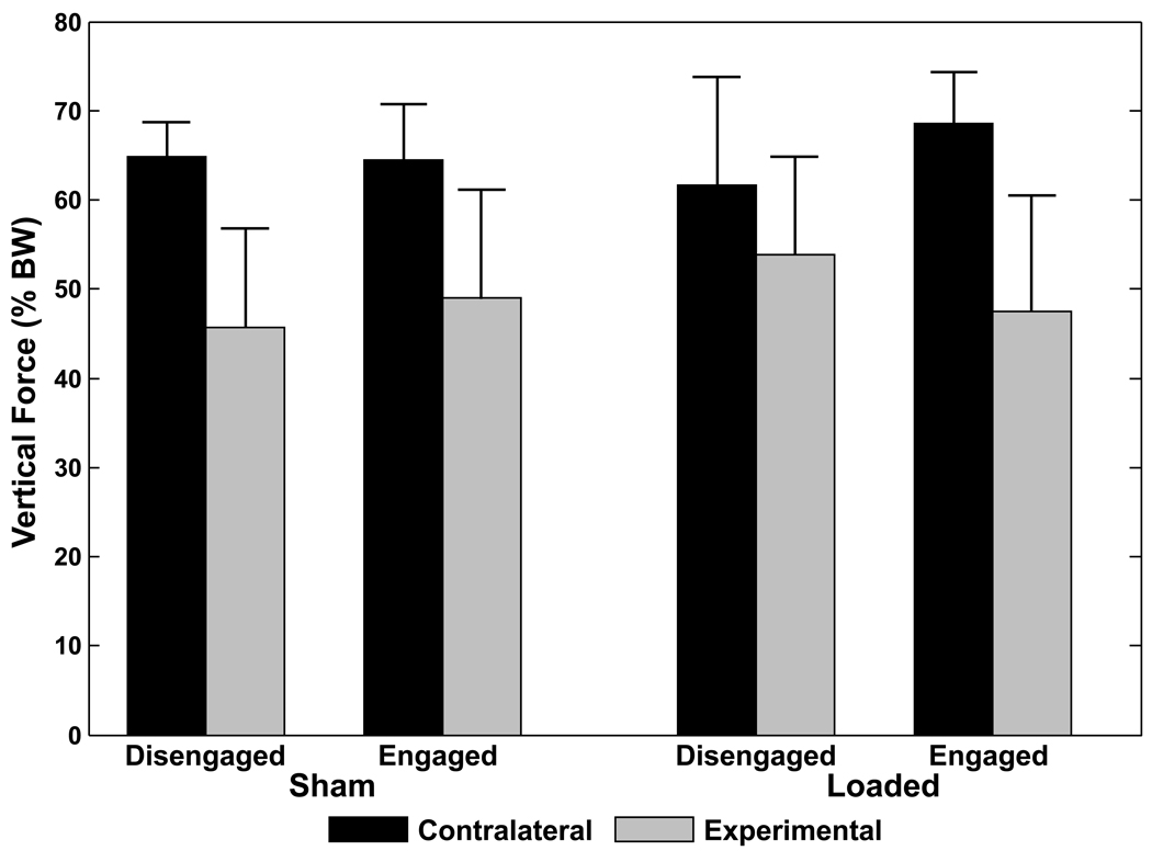
Comparisons across Sham and Loaded groups to examine the effects of applying the compressive overload to the medial compartment with the VLD found no significant differences between the VLD being Disengaged or Engaged or between Sham and Loaded groups on any gait measures (Figure 4; Table 3). Independent of VLD status (Disengaged, Engaged) and group (Sham, Loaded), the Contralateral and Experimental legs were significantly different from each other for all gait measures except contact time which replicates the surgical effects found in the initial set of analyses (Table 3).
Figure 4.
Mean peak vertical forces as a percent of body weight (% BW) for contralateral and experimental legs for both the Sham and Loaded groups with the varus loading device (VLD) disengaged (no compressive overload applied) and engaged (compressive overload applied) (Mean +/− SEM). Statistical analyses found a significant effect of leg (p=0.02). There were no statistical differences between the Sham and Loaded groups nor between VLD disengaged or engaged.
Table 3.
Gait measures (mean ±SD) for experimental and contralateral legs in Loaded and Sham groups with the varus loading device (VLD) engaged (compressive overload applied) and disengaged (no compressive overload applied). There were no significant differences between the Loaded and Sham groups and no significant differences between the VLD being engaged and disengaged for the Loaded or Sham groups.
| Sham Group |
Loaded Group |
|||||||
|---|---|---|---|---|---|---|---|---|
| Contralateral Leg |
Experimental Leg |
Contralateral Leg |
Experimental Leg |
|||||
| Gait Parameter |
Engaged |
Disengaged |
Engaged |
Disengaged |
Engaged |
Disengaged |
Engaged |
Disengaged |
| Contact Time (ms) | 502±177 | 548±104 | 379±42 | 441±83 | 466±149 | 500±169 | 450±69 | 472±14 |
| Braking Force (% BW)* | −7.4±3.7 | −5.8±2.3 | −3.0±2.8 | −4.4±2.8 | −5.8±3.5 | −8.7±3.1 | −3.9±2.4 | −4.6±1.8 |
| Propulsive Force (% BW)* | 9.9±4.9 | 10.4±1.4 | 5.4±2.5 | 4.7±2.3 | 11.0±1.6 | 9.5±2.4 | 10.5±12.2 | 6.8±2.3 |
| Medial Force (% BW)* | −6.8±2.7 | −8.1±1.8 | −2.1±1.9 | −2.1±0.8 | −6.6±0.9 | −6.8±1.6 | −3.1±2.0 | −4.6±2.6 |
| Lateral Force (% BW)* | 2.1±1.3 | 1.9±1.0 | 8.2±2.8 | 7.9±2.5 | 1.4±0.4 | 2.0±1.3 | 5.2±2.8 | 5.3±.4.0 |
| Vertical Force (% BW)* | 64.5±6.2 | 64.8±3.8 | 49.0±12.3 | 45.7±11.1 | 68.6±5.7 | 61.7±12.2 | 47.5±13.0 | 53.9±11.0 |
Significant effect of Leg (Contralateral vs. Experimental) independent of Group or Engagement status (p<0.01).
4. DISCUSSION
Evaluation of gait measures during spontaneous walking found the surgery and attachment of the VLD significantly changed the gait in the experimental leg. No additional gait alterations occurred with engagement of the VLD.
The bone plates were implanted without disrupting the integrity of the musculature, joint capsule, ligaments/tendons, vasculature or nerves. Stretching of surrounding tissues to expose the bone, bleeding from drilling into the bone, or elicitation of inflammation may have contributed to the surgical effect observed in gait parameters. Since gross cartilage erosion was not observed in the tibiofemoral joints in any of these rats at necropsy (following 12 weeks of altered loading), the observed gait alterations were attributed to the surgical procedure and device attachment rather than degenerative joint changes.
The VLD and bone plates weigh 0.1 N which is less than 2% of the weight of the rats. This mass was estimated to be ~15–19% of the mass of the rear leg of the Sprague-Dawley rats. The VLD mass compared to the rear leg mass may explain the shift in the mediolateral force from the medial to lateral direction (Table 2) since the VLD is attached to the lateral aspect of the femur. However, the VLD mass should have caused an increase and not the observed decrease in vertical force.
Although the normal distribution of forces between the medial and lateral compartments of the rat knee is not yet known, the overall loading experienced in the medial compartment of the knee with engagement of the VLD is a summation of normal loads produced by body weight and muscle contraction plus the applied static overload (50–80% BW) (Roemhildt et al., 2010, (Roemhildt, 2009). While a reduction in vertical ground reaction force was observed with the attachment of the VLD (17–30% BW for Sham and Loaded groups), this was a small proportion of the applied compressive overloads. Therefore, the ground reaction forces and those applied by the VLD combined to produce a net increase in in vivo loads in the tibiofemoral joint.
The rear foot transmits most of the vertical ground reaction forces in the walking rat (Clarke, 1995). In a rat model of injection-induced rapid arthrosis, a reduction in peak vertical force was observed in the experimental leg with the contralateral fore leg transmitting the extra load (Clarke et al., 1997). The forces in the fore leg were not measured in the present study, however the observation that the forces in the contralateral rear leg were unchanged while a decrease in vertical reaction force was observed in the experimental rear leg suggests the extra load was shifted to the fore legs. Gait data for rat transection models of osteoarthritis is not yet available barring comparison of the effects of surgery from these models with those observed with the VLD model.
Although animals appeared to ambulate normally following their 2-week recovery, reduced vertical ground reaction force was observed in the experimental limb. These alterations may be related to limping, however, there was no evidence of increased force in the contralateral limb nor differences in contact times between legs as is typical in quadrupedal animal limping (Bockstahler et al., 2007; Suter et al., 1998). This study was not designed to investigate sources of gait alterations such as pain.
Pre-surgical baseline measures were not collected in this study nor were adaptations to the device over time evaluated. The findings here at 12–14 weeks post-operatively suggest that future studies could investigate the time course of gait alteration.
The significant reduction in vertical ground reaction force due to the surgical attachment of the VLD affects the resulting contact stresses observed in the tibiofemoral joint and must be considered when interpreting the results of studies which use this model to study the effects of altered loading on cartilage biomechanics and biology. No additional changes in gait measures were observed with application of medial compartment compressive overload suggesting that Sham animals can be used as a control for animals with compressive overload.
Supplementary Material
ACKNOWLEDGEMENTS
The authors are grateful to Dr. Nelson Tacy, Amy Gassman, and Danielle Funaro for their assistance with the rat care and procedures and Synthes for providing the bone screws used in this work. Funded by NIH AR052815.
Footnotes
Publisher's Disclaimer: This is a PDF file of an unedited manuscript that has been accepted for publication. As a service to our customers we are providing this early version of the manuscript. The manuscript will undergo copyediting, typesetting, and review of the resulting proof before it is published in its final citable form. Please note that during the production process errors may be discovered which could affect the content, and all legal disclaimers that apply to the journal pertain.
CONFLICT OF INTEREST STATEMENT
The authors have no commercial or other relationships which may lead to a conflict of interest.
REFERENCES
- 1.Bockstahler BA, Skalicky M, Peham C, Müller M, Lorinson D. Reliability of ground reaction forces measured on a treadmill system in healthy dogs. The Veterinary Journal. 2007;173(2):373–378. doi: 10.1016/j.tvjl.2005.10.004. [DOI] [PubMed] [Google Scholar]
- 2.Clarke KA. Differential fore- and hindpaw force transmission in the walking rat. Physiology & Behavior. 1995;58(3):415–419. doi: 10.1016/0031-9384(95)00072-q. [DOI] [PubMed] [Google Scholar]
- 3.Clarke KA, Heitmeyer SA, Smith AG, Taiwo YO. Gait Analysis in a Rat Model of Osteoarthrosis. Physiology & Behavior. 1997;62(5):951–954. doi: 10.1016/s0031-9384(97)00022-x. [DOI] [PubMed] [Google Scholar]
- 4.O'Connor BL, Visco DM, Heck DA, Myers SL, Brandt KD. Gait alterations in dogs after transection of the anterior cruciate ligament. Arthritis & Rheumatism. 1989;32(9):1142–1147. doi: 10.1002/anr.1780320913. [DOI] [PubMed] [Google Scholar]
- 5.Roemhildt ML, Fleming BC, Beynnon BD, Churchill D. In Vitro Validation of a Varus Loading Device in the Rabbit Knee. Proceedings of the World Congress on Osteoarthritis and cartilage Montreal, QC. 2009 [Google Scholar]
- 6.Suter E, Herzog W, Leonard TR, Nguyen H. One-year changes in hind limb kinematics, ground reaction forces and knee stability in an experimental model of osteoarthritis. Journal of Biomechanics. 1998;31(6):511–517. doi: 10.1016/s0021-9290(98)00041-4. [DOI] [PubMed] [Google Scholar]
- 7.Vilensky JA, O'Connor BL, Brandt KD, Dunn EA, Rogers PI, Delong CA. Serial kinematic analysis of the unstable knee after transection of the anterior cruciate ligament: Temporal and angular changes in a canine model of osteoarthritis. Journal of Orthopaedic Research. 1994;12(2):229–237. doi: 10.1002/jor.1100120212. [DOI] [PubMed] [Google Scholar]
- 8.Zumwalt AC, Hamrick M, Schmitt D. Force plate for measuring the ground reaction forces in small animal locomotion. Journal of Biomechanics. 2006;39(15):2877–2881. doi: 10.1016/j.jbiomech.2005.10.006. [DOI] [PubMed] [Google Scholar]
Associated Data
This section collects any data citations, data availability statements, or supplementary materials included in this article.


