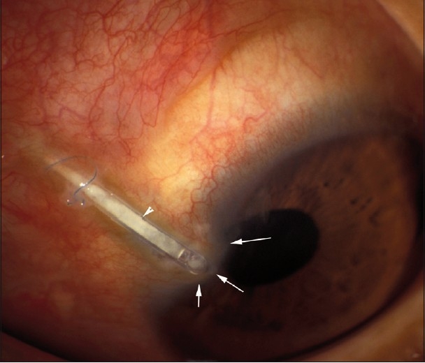
Figure 1.

Photomicrograph demonstrating transconjunctival (arrowhead) and corneal erosion of the tube as evidenced by corneal thinning and full thickness opacity (arrows) surrounded by hyperemic conjunctiva. Note the broken prolene anchoring suture at the site of conjunctival erosion
