Figure 2.

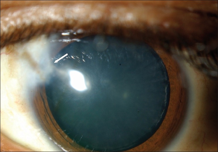
The anterior segment of the left eye following removal of the extruded tube. A triangular corneal opacity in the area of erosion is seen (arrow)

The anterior segment of the left eye following removal of the extruded tube. A triangular corneal opacity in the area of erosion is seen (arrow)