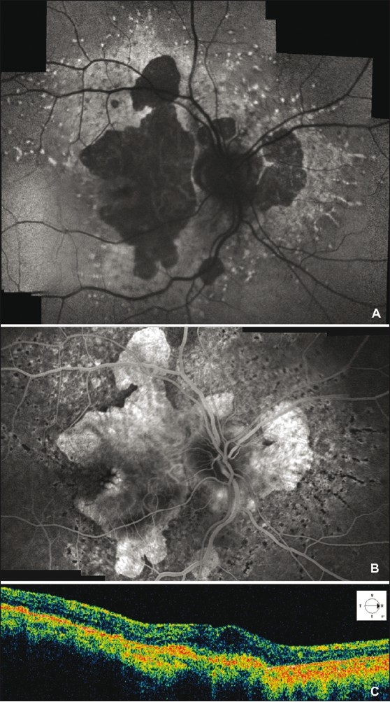
Figure 2.

Fundus autofluorescence shows retinal flecks and areas of well-defined retinal atrophy 1 month after intravitreal bevacizumab injection (A). Fluorescein angiography late frame of the right eye, one month after intravitreal bevacizumab injection, shows moderate heterogeneous hyperfluorescence and absence of leakage due to the choroidal neovascularization (CNV) closure (B). Optical coherence tomography scan demonstrates high reflective mass corresponding to the subfoveal fibrotic CNV, and absence of both neurosensory retina elevation and cystoid macular edema in the macular area (C)
