Figure 1.
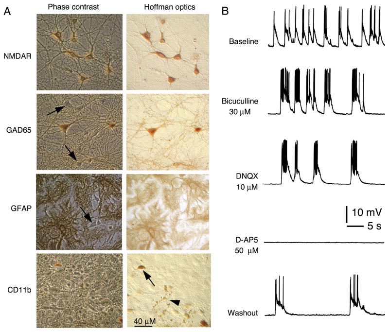
Hippocampal cultures. A. Digitized images of immunostained cultures showing cell types in the hippocampal cultures. The phase contrast images show all cells in the field while the Hoffman optics images show the immunostained cells. The well-developed hippocampal cultures contain excitatory and inhibitory neurons and glial cells, thus reflecting the hippocampus in vivo. The neurons can be identified by their distinct morphological characteristics and by immunostaining for specific proteins. The majority of hippocampal neurons express NMDAR1. Inhibitory neurons can be identified by immunostaining with an antibody to glutamic acid decarboxylase (GAD65), the synthetic enzyme for gamma amino butryic acid (GABA) the transmitter used by the inhibitory neurons. In the phase contrast image, arrows point to some neurons that are unstained for GAD65. These neurons are presumably excitatory neurons that use glutamate as a transmitter. The primary glial cell type in culture is the astrocyte, which can be identified by immunostaining for the astrocyte specific protein GFAP. In the phase contrast image, the arrow points to a neuron, which is unstained for GFAP. Some microglia are also present in culture and can be identified by immunostaining for the microglial marker CD11b. Arrow in the Hoffman optics image points to a microglia; arrow head points to immunostained processes from another microglia. B. Patch clamp recordings from a cultured hippocampal neuron showing the spontaneous synaptic network activity. The activity consists of synaptic potentials that evoke action potentials. The contribution of inhibitory (GABA-mediated) and excitatory (glutamate-mediated) synaptic events to the activity can be shown by application of GABA and glutamate receptor antagonists. The recordings were made in physiological saline with reduced Mg2+ levels (30 μM) to remove Mg2+ block of NMDARs. In the reduced Mg2+ saline, NMDAR-mediated synaptic responses can be observed at resting membrane potential. Changes in the spontaneous network activity with various receptor antagonists show that GABA-A, AMPA and NMDA receptors play a central role in generating the synaptic network activity. Bicuculline blocked the GABA-A receptors that mediate inhibitory synaptic responses, whereas AMPA and NMDA receptor-mediated excitatory synaptic responses were blocked by DNQX and D-AP5, respectively.

