Figure 1.
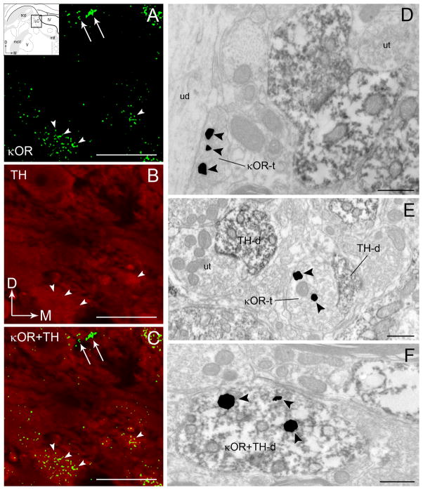
A–C. Confocal fluorescence photomicrographs showing κOR and tyrosine hydroxylase (TH) immunoreactivities in the locus coeruleus (LC). κOR immunoreactivity was labeled with fluorescein isothiocyanate (green) and TH was labeled with rhodamine isothiocyanate (red). Arrowheads point to κOR-labeled processes that are localized in TH-labeled perikarya that can also be seen in the merged image in panel C. Arrows point to varicose processes that only contain κOR that are also seen in the merged image in panel C. Arrows indicate the dorsal (D) and medial (M) orientation of the tissue section. Inset shows schematic diagrams adapted from the rat brain atlas of Swanson (1992) depicting the LC region sampled. In the insets, arrows indicate dorsal (D) and medial (M) orientation of the sections illustrated. Abbreviations: scp, superior cerebellar peduncle; IV, fourth ventricle; mlf, medial longitudinal fasciculus; moV, motor root of the trigeminal nucleus; V, motor nucleus of the trigeminal nucleus. D–F. Electron photomicrographs showing immunoperoxidase labeling for TH and immunogold-silver labeling for κOR in the LC. D. An immunoperoxidase-labeled TH dendrite and an immunogold-silver labeled (arrowheads) κOR terminal (κOR-t) are seen in the same field. κOR-t targets an unlabeled dendrite (ud). Located nearby is an unlabeled axon terminal (ut) targeting a TH-d. E. An immunogold-silver labeled (arrowheads) κOR-t is shown contacting a TH-d. In the same field is shown an unlabeled terminal (ut) contacting a TH-d. F. A TH-d exhibiting immnoperoxidase labeling also exhibits immunogold-silver labeling (arrowheads) for κOR (κOR+TH-d). Scale bars, 100 μm (A-C), 0.5μm. (D–F).

