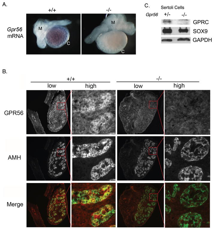
Figure 1. Gpr56 mRNA and its encoded protein are expressed in Sertoli cells comprising the testis cords of mouse embryos.
A. The distribution patterns of Gpr56 mRNA in the genital ridges from E16.5 Gpr56+/+ (n = 3) and Gpr56−/− (n = 3) embryos were examined by in situ hybridization using Gpr56 antisense probe. The probe specifically detected Gpr56 mRNA in wild-type but not in testis cords from Gpr56−/− mice. Gpr56−/− gonads often appear larger than Gpr56+/− gonads of the same stage. The reason is not known. M: mesonephros; C: coelomic epithelium.
B. The distribution pattern of GPR56 protein in the genital ridges from E14.5 Gpr56+/+ (n = 4) and Gpr56−/− (n = 4) embryos was examined by co-immunostaining with the anti-GPR56 antibody (red) and anti-AMH antibody (green), which labels Sertoli cells. Colocalization of GPR56 and AMH was observed in the wild-type but not in the Gpr56−/− gonads. Images of whole genital ridges are shown in low magnification on the left, with an area in each gonad (red box) shown in high magnification on the right.
C. GPR56 was specifically detected in the Gpr56+/− Sertoli cell preparations. Sertoli cells were isolated from two 4-week old Gpr56+/− and Gpr56−/− mice, lysed, and probed with the anti-GPR56 antibody and the anti-SOX9 antibody on a western blot. The level of GAPDH in each lysate was used as the loading control.

