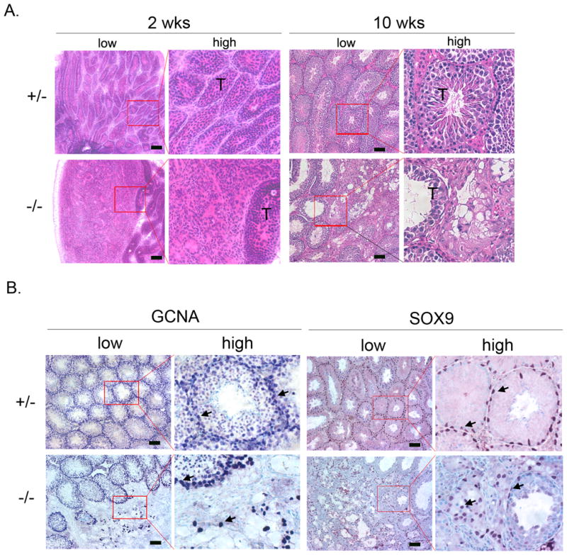
Figure 3. Disruption of seminiferous tubule formation in Gpr56−/− testes.

A. H&E staining of testis sections from 2-week old (n = 3) or 10-week old (n > 8) Gpr56+/− and Gpr56−/− mice. A disruption of seminiferous tubules (T) in Gpr56−/− sections is apparent at both stages. Images of low magnifications are shown on the left, with some areas (red box) shown in high magnifications on the right.
B. The distribution of germ cells and Sertoli cells are abnormal in the Gpr56−/− testes. Sections of testes from 10-week old Gpr56+/− (n = 4) and Gpr56−/− mice (n = 4) were stained with the anti-GCNA1 antibody to detect germ cells (arrows) and the anti-SOX9 antibody to detect Sertoli cells (arrows). Images of low magnifications are shown on the left, with some areas (red box) shown in high magnifications on the right. Depletion of germ cells and disorganized localization of Sertoli cells are seen in the disrupted areas of the Gpr56−/− testes. Scale bar: 50 μm (A, B).
