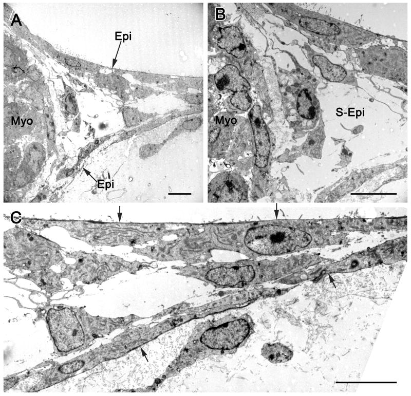
Figure 1.
Transmission electron micrographs illustrating epicardial cell formation of vascular tubes in a collagen matrix. In A, the edge of the embryonic mouse heart explant with myocardium (myo) is seen along with epicardial (epi) cells emerging from the explant. The region between the emerging epicardial cells is an extension of the subepicardium. A detail of this micrograph (B) shows subepicardial cells (S-Epi) in the lumen of the vascular tube. In C, the components of vascular tube distal to the explant are seen, with epicardial cells indicated by arrows. The lumen, containing subepicardial cells (S-Epi), is between the epicardial cells, which by immunohistochemistry are positive for the endothelial cell marker BS-I. These images are typical of tube formation in both mouse and quail explants, i.e., epicardial cells delaminate and form the walls of the tubes.

