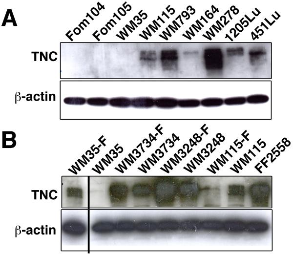
Figure 1. TNC expression is up-regulated in melanoma cells.
(A) Immunoblot analysis to assess levels of TNC in human melanocytes and in melanoma cell lines. Increased levels of TNC in multiple melanoma cell lines as compared to normal melanocytes. Foreskin melanocytes: Fom 104 and Fom 105; melanoma cell lines: WM35, WM115, WM793, WM164, WM278, 1205Lu, and 451Lu. (B) TNC expression in either sphere-forming melanoma cells or adherent melanoma cells. Increased levels of TNC in sphere-forming melanoma cells (denoted with a –F suffix) as compared to adherent melanoma cells. Melanoma cell lines: WM35, WM3734, WM3248 and WM115; fibroblasts: FF2558. β-actin immunoblotting was performed as a loading control. The furthermost left lane was originally the last lane; the location was switched for clearer comparison of WM35-F and WM35.

