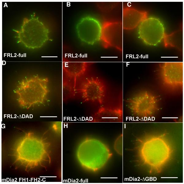
Figure 8. Full-length FRL2 has reduced ability to assemble filopodia.
A-F) Jurkat cells were transfected with un-tagged constructs containing either full-length FRL2 (A-C), or FRL2 DDAD (D-F). After 6 hrs, cells were plated onto poly-lysine for 10 min, fixed, and stained with rhodamine-phalloidin (red) and anti-FRL2 followed by fluorescently labeled secondary antibody (green). Three examples of transfected cells are shown for each FRL2 construct. A) shows an example of a full-length FRL2 cell scored “positive” for filopodia, whereas B) and C) are “negative”. D)-F) are all examples of “positive” cells transfected with FRL2 ΔDAD. G-H) Jurkat cells transfected with GFP-fusions of mDia2 constructs. G) is mDia2 FH1-FH2-C, H) is mDia2 full-length, I) is mDia2-ΔGBD. Scale bars represent 5 μm.

