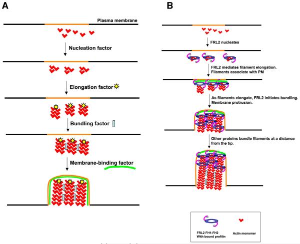
Figure 9. Filopodia assembly models.
A) Schematic representations of four biochemical activities needed for filopodia assembly from a designated region of the plasma membrane (orange). Theoretically, membrane binding could occur at any step, though we include it as the last step. Also, membrane binding could occur at the tips and/or the sides of filopodia.
B) Working model for FRL2 FH1-FH2-mediated filopodia assembly, taking into account its known biochemical activities of nucleation acceleration, elongation regulation, and bundling. FRL2 FH1-FH2 nucleates filaments. Barbed end-bound FRL2 FH1-FH2 facilitates filament elongation through profilin. Additional FRL2 molecules mediate bundling near the tip. As the filaments elongate, other proteins bundle the filaments further from the tip. An unknown factor mediates membrane association. We depict membrane association at the elongation step, but it could occur during another step.

