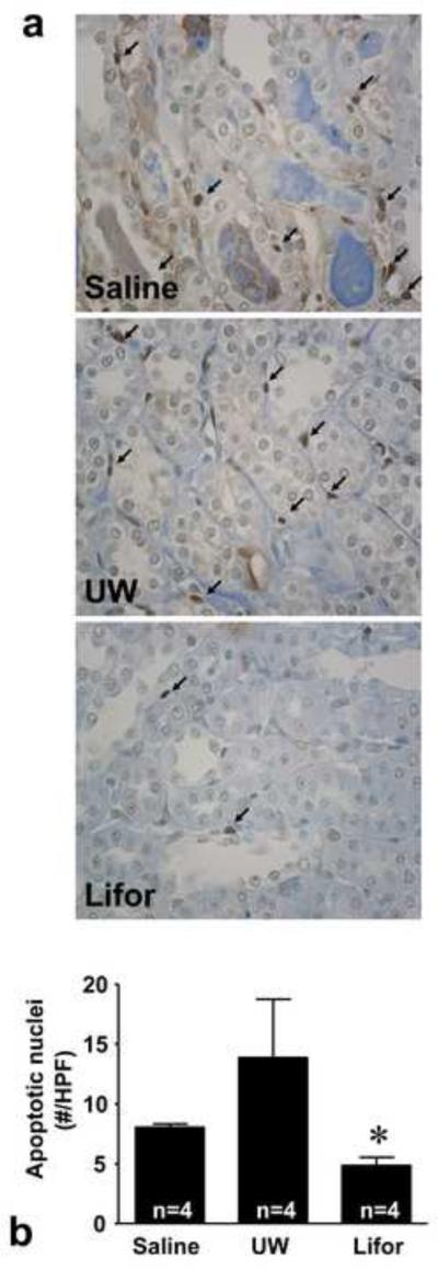
Figure 3. Effect of Lifor on apoptosis 24 hours after warm renal IR injury.
Apoptosis was assessed by TUNEL staining. (a) Representative photomicrographs of TUNEL staining depicting apoptotic nuclei (arrows) in saline, UW, and Lifor perfused kidneys. Apoptotic nuclei were detected in renal tubular epithelial cells and peri-tubular interstitial cells. (b) The number of apoptotic nuclei was significantly less in rat kidneys perfused with Lifor compared to saline or UW solution. N=4 animals/group. At least 10 fields per section and 2 sections per animal were counted. Mean values ± SE are presented. * P < 0.05 vs. saline group.

