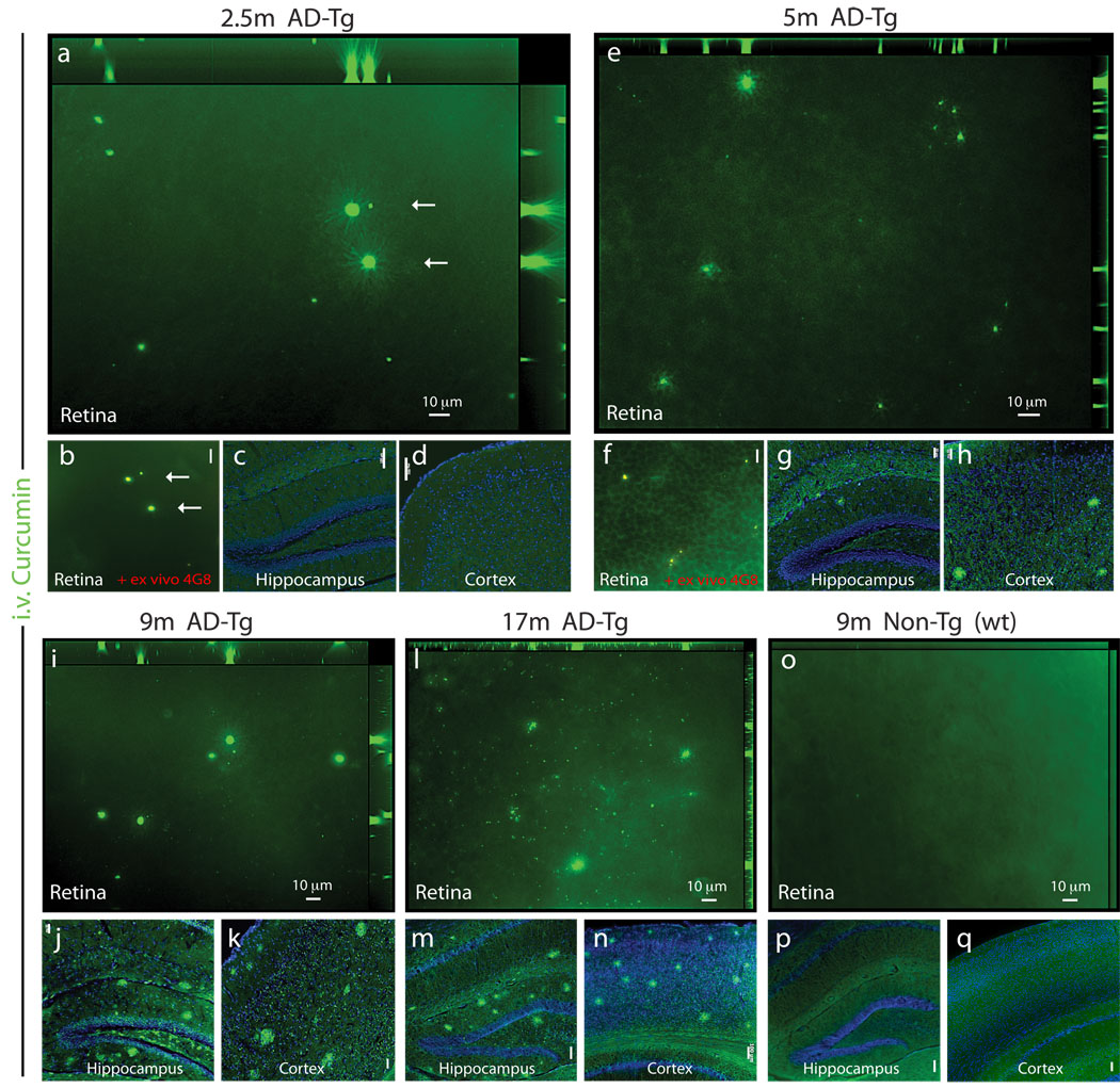
Figure 2.
Bioavailability of systemically administered curcumin to the mouse eye: accumulation of curcumin-labeled retinal Aβ plaques with disease progression and detection at early pre-symptomatic stage. (a–q) Representative z-axis projection images of whole-mount retinas and brain coronal cryosections from AD-Tg and non-Tg (wt) mice at various ages, following i.v. curcumin injections for 5 days: In 2.5-month-old AD-Tg mouse (a,b) retinal curcumin-labeled in vivo of Aβ plaques (green) were visible, with further (b) validation of Aβ plaque identity in the same retinal location (arrows) by staining ex vivo with anti-human Aβ mAb 4G8 (secondary Ab-Cy5 conjugate). Co-localization of curcumin and Aβ antibody in yellow pseudocolor). Scale bar = 10 µm. (c,d) No plaques were detected in the brain hippocampus and cortex of 2.5-month-old AD-Tg mice. Scale bars = 100 µm. (e–h) 5-month-old AD-Tg mouse: (e) Presence of curcumin-labeled plaques in the retina and (f) co-labeling following 4G8 mAb staining ex vivo. Scale bar = 20 µm; (g,h) detection of plaques in the brain. Scale bars = 50 µm. (i–k) 9-month-old AD-Tg mouse: (i) Multiple plaques in the retina and (j,k) in the brain. Scale bars = 50 µm. (l–n) 17-month-old AD-Tg mouse: (l) Numerous plaques in the retina and (m,n) in the brain. Scale bars =100 µm. (o–q) 9-month-old non-Tg (wt) mouse: (o) No Aβ plaques in the retina nor (p,q) in the brain. Scale bars =100 µm.

